
Professor of Rhinology & Skull Base Surgery, Ain Shams University, Cairo, Egypt
Department of Oto-Rhino-Laryngology, Faculty of Medicine, Ain Shams University, Cairo, Egypt.
14- Endoscopic Nasal Surgery For Maxillary Atelectasis
Chronic Maxillary Atelectasis
Chronic Maxillary Atelectasis (CMA) is characterized by a reduced maxillary sinus volume due to an inward bowing of one or more of the sinus walls.
The silent sinus syndrome represents maxillary sinus atelectasis that results in painless enophthalmos, hypoglobus and facial asymmetry. Some authors restrict the term to patients with no history of sinusitis, trauma or surgery.
Clinical Presentation:
Most patients were symptomatic, and some presented with relatively long-standing facial asymmetry, enophthalmos of 2-5 mm, hypoglobus and diplopia. Symptoms of sinusitis are not always present. Where there is significant deformity of the orbital floor, patients may develop diplopia. However, extraocular movements are usually normal.
Imaging Studies:
Inward bowing of the antral walls and persistent opacification on CT is diagnostic.
Pathogenesis:
The uncinate process is thinned and retracted to the orbital wall, obstructing the maxillary sinus infundibulum. The medial infundibular wall causes valvular occlusion of the natural ostium resulting in intrasinus negative pressure (-8.4 2.6 cm water) that in turn results in gradual inward bowing of all four of the maxillary walls: roof (orbital floor), medial, posterolateral and anterior walls. Orbital volume increases with resultant enophthalmos, hypoglobus with lowering of the orbital floor and variable flattening of the malar eminence.
Treatment and prognosis
The condition is benign but may result in cosmetic deformity and diplopia. Treatment involves middle meatal antrostomy. Once drainage is established, no further volume loss will develop. However, any deformity at the time of surgery will be permanent and may require surgical correction, especially if diplopia is present, using a combination of bone allograft and porous polyethylene sheets.
Video Demos Of Interesting Case:
Movie 1:
28 y.o. female patient with slowly-developing left enophthalmos and hypoglobus for
5 months. CT study done in Jan., 2011 showed left maxillary atelectasis with the
left middle turbinate touching the lateral nasal wall and intramaxillary partial
fluid opacity.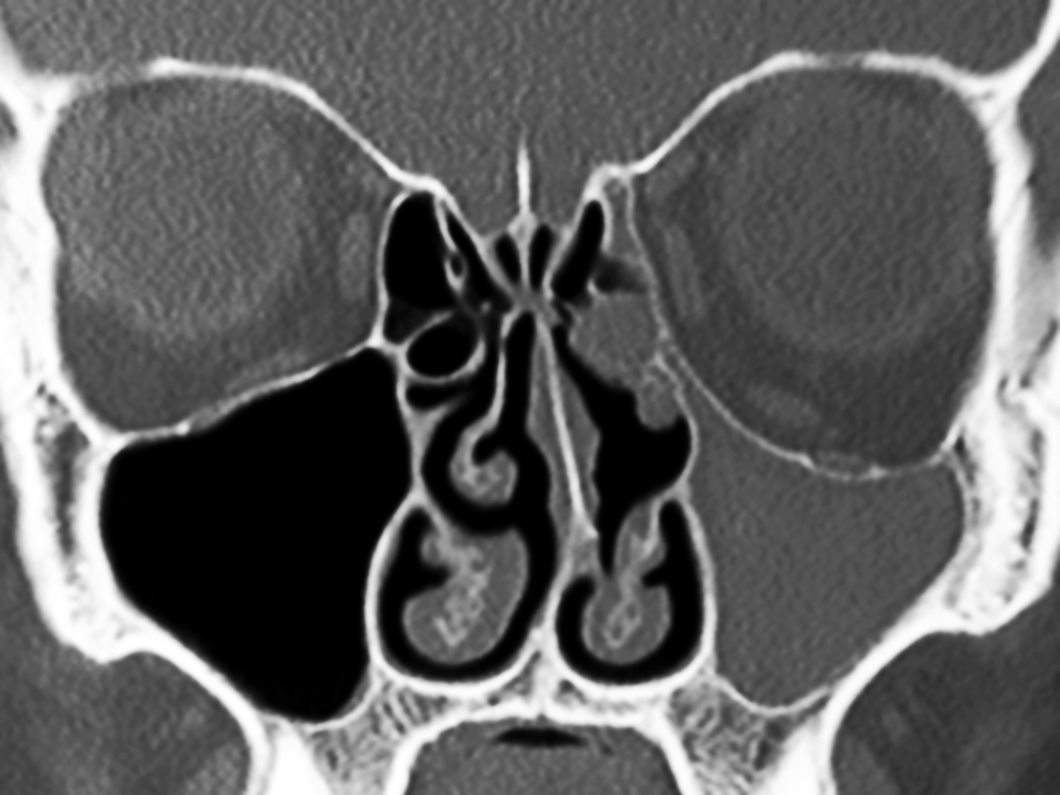 A second study done in March, 2011 showed the same collapse of the middle
meatus, ethmoidal infundibulum and total opacity of the atelectatic maxillary
sinus beside opacification of the left anterior ethmoid and left frontal
sinuses.
A second study done in March, 2011 showed the same collapse of the middle
meatus, ethmoidal infundibulum and total opacity of the atelectatic maxillary
sinus beside opacification of the left anterior ethmoid and left frontal
sinuses.
Endoscopic Sinonasal Surgery For Maxillary Atelectasis Movie
"It is high resolution video, about 100 MB in size, better download it first: Right-Click>Save Target as in Windows & Download Linked File in Mac"